
Two confocal Raman spectroscopic analysis systems are installed in the laboratory, in addition to integrated atomic force microscopy (AFM) with an AIST-NT scanning probe microscope, which also allows the use of tip-enhanced Raman spectroscopy (TERS).
- LabRAM 300, Horiba (formerly Jobin Yvon), acquired in 1998.
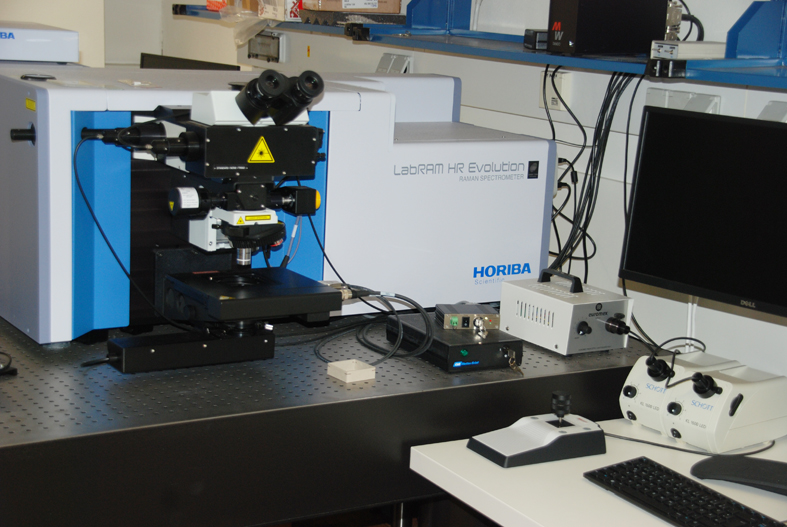
- LabRAM HR Evolution 800, Horiba, acquired in 2018
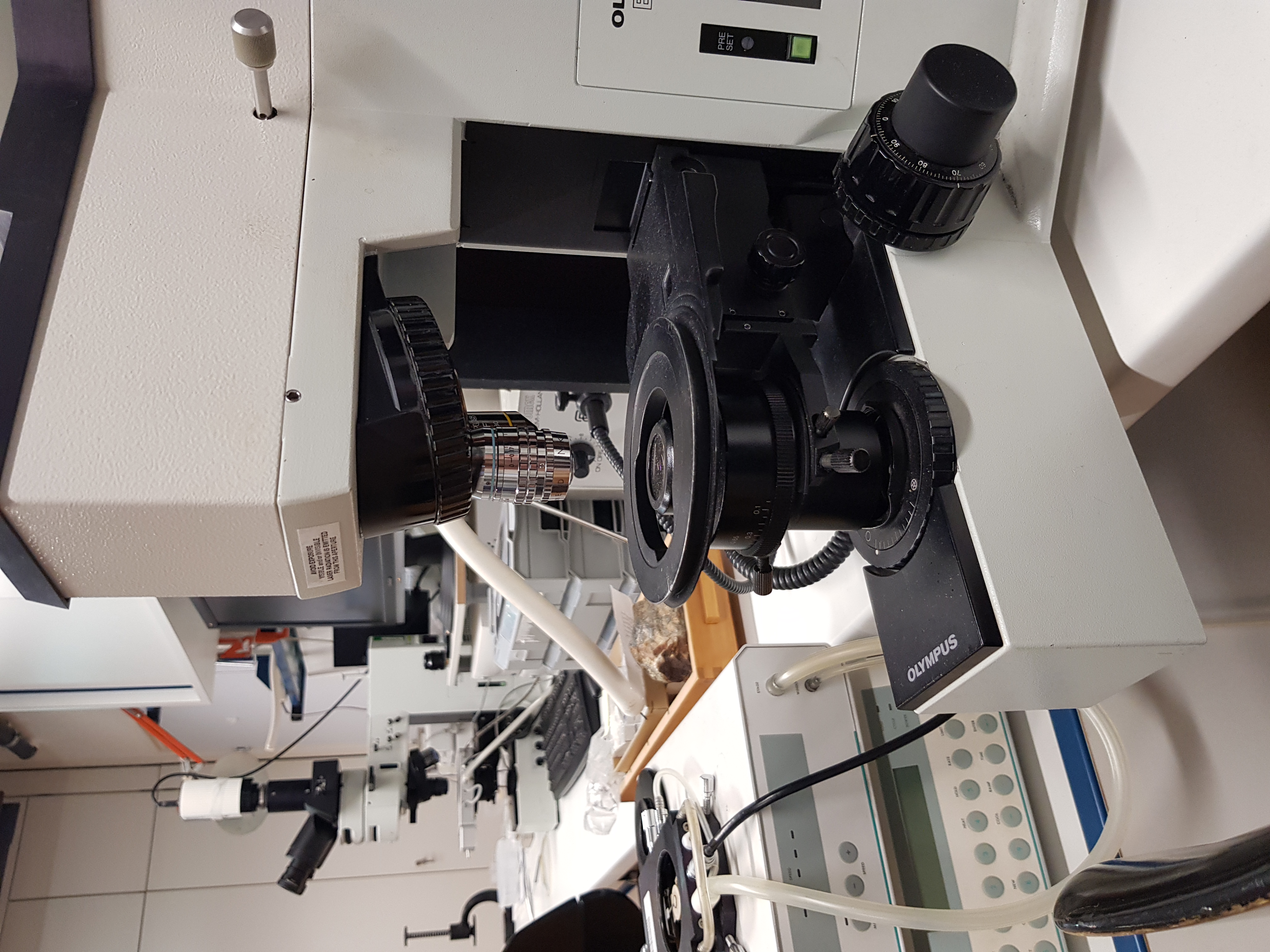
LabRAM 300
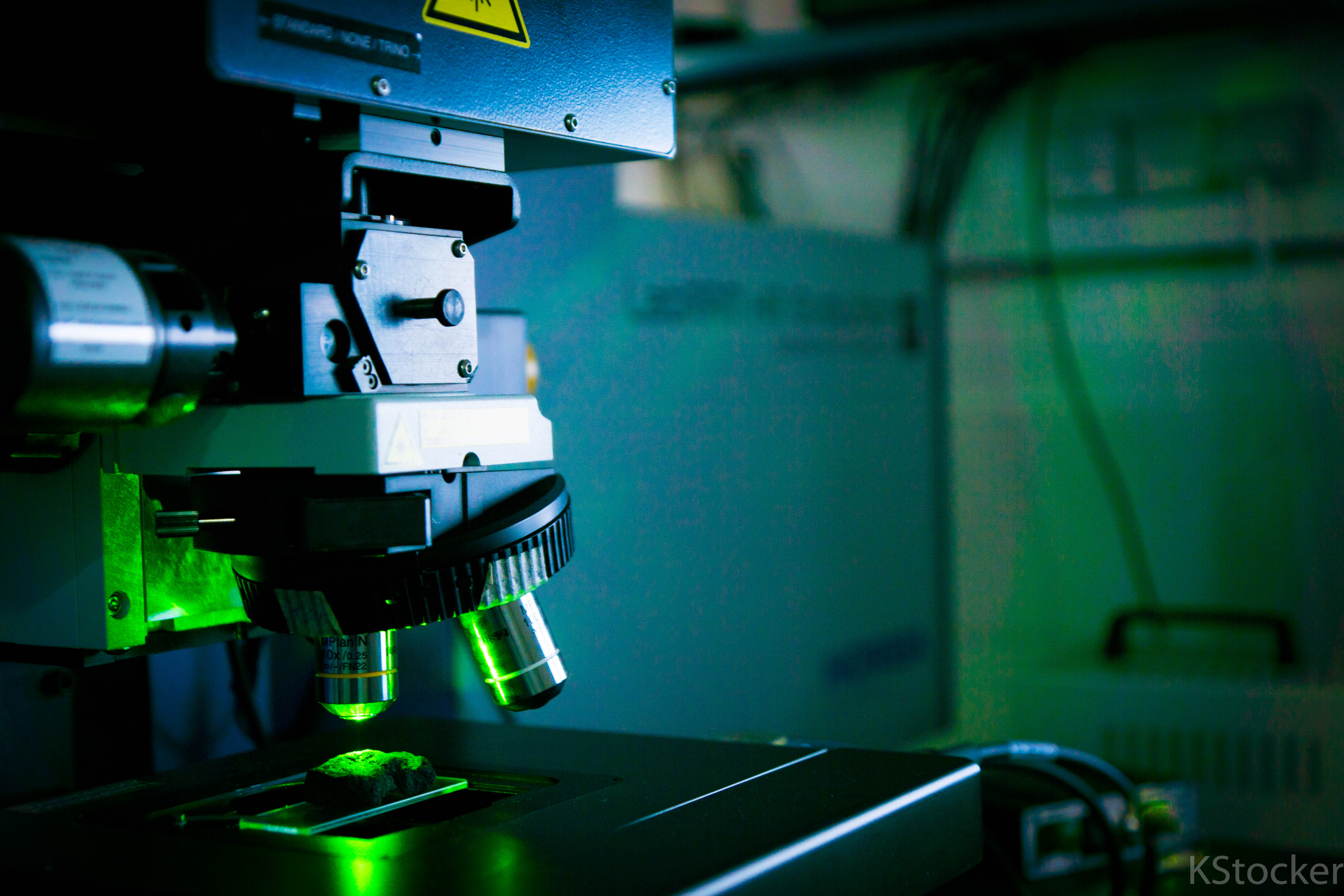
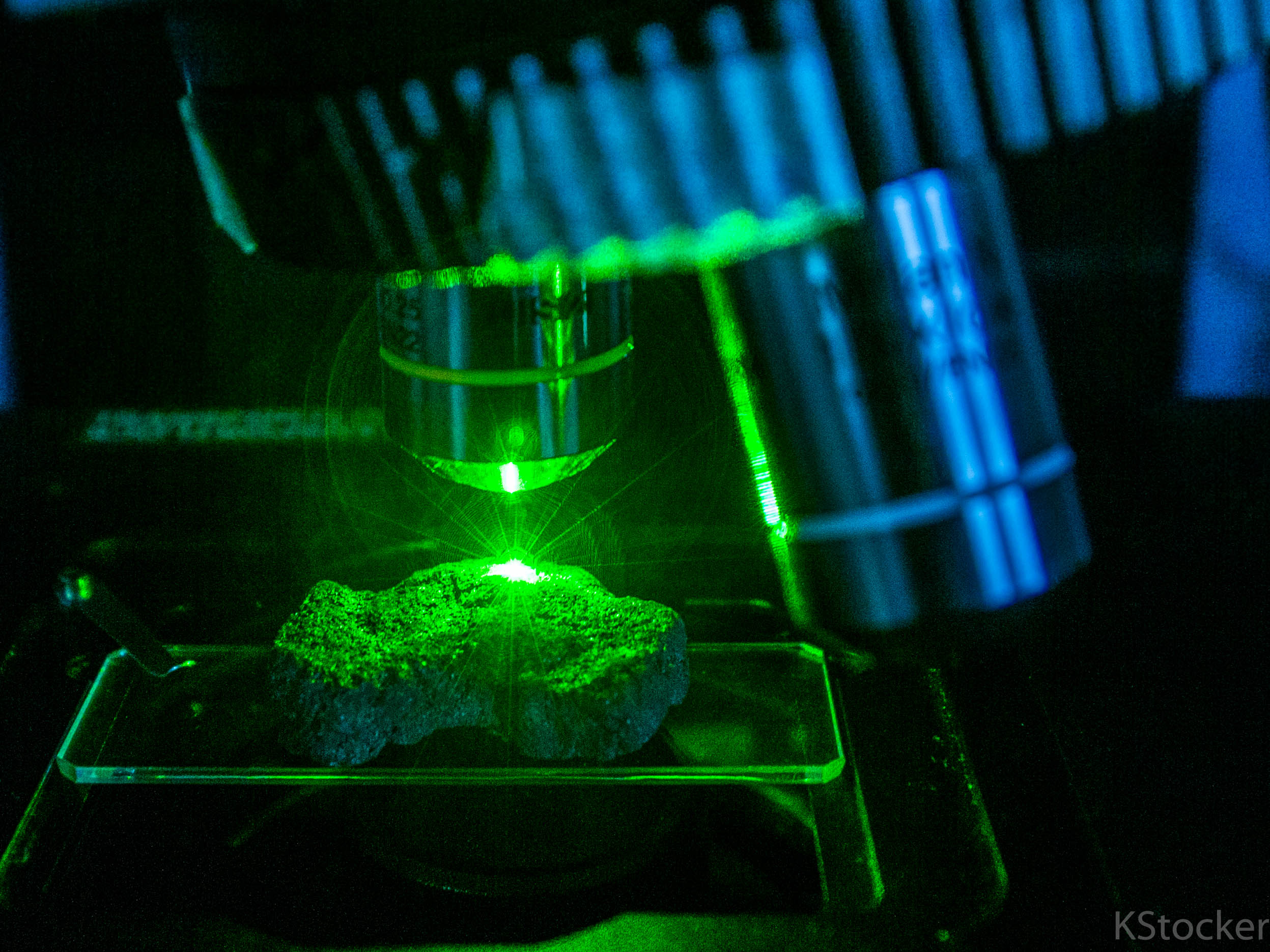
Frequency doubled Nd-YAG laser (100 mW, 532.2 nm); diffraction grating with 600 and 1800 grooves/mm; detection with a Peltier-cooled CCD matrix detector with slow scanning; Laser focusing and sample viewing with a simplified Olympus microscope (without eyepiece) equipped with various objectives (5x, 10x, 40x, 50x, 80x, 100x) with observation in reflected and transmitted light; the 40x and 100x objectives are long working distance objectives used for liquid inclusion research. LabSpec 5 software is used to control the analysis settings.
A mobile Linkam heating freezing stage (THMSG 600) can be connected to the system to enable Raman measurements at temperatures between -190 ˚C and 600˚C.
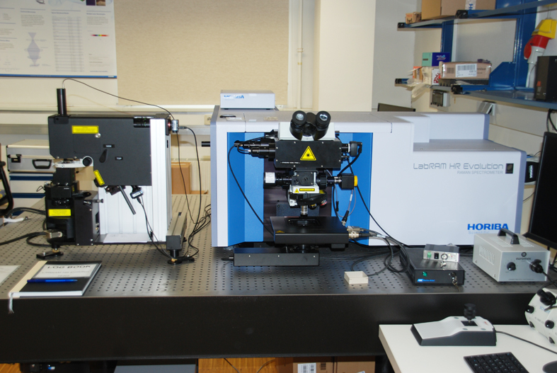
LabRAM HR Evolution
High resolution confocal Raman microscope and scanning probe microscope; with fully integrated solution for nanospectroscopy (AFM-Raman, TERS and SNOM); 800 mm Czerny-Turner spectrograph; Nd-YAG laser (532 nm), He-Ne laser (632,816 nm); integrated Olympus BX-FM open microscope. 5x, 10x and 100 x objectives; 9 density filters (wheel); holder for interference filters; edge filters for Rayleigh and anti-Stokes scattering; 600, 1800 and 2400 grating; Synapse EM detector, 1600x200 pixels, open electrode chip, Peltier-cooled -60 ˚C, pixel size 16x16 µm; motorised XYZ microscope stage, polarisation accessories; software package LabSpec 6, KnowItAll database.
AIST SmartSPM
AFM applications: Contact, semi-contact, non-contact, lateral force microscopy (LFM), piezoelectric force microscopy (PFM), phase contrast, magnetic force microscopy (MFM), single-pass MFM, electrostatic force microscopy (EFM), single-pass EFM, atomic force microscopy (SCM), SCM; flexibly guided high-speed SPM XYZ scanner with a scanning area of 100 x 100 x 15μm; SPM platform with manual XY stage, 25 x 25 mm; fully automatic alignment of AFM feedback laser and AFM photodiode sensor, no manual intervention required; fully automatic alignment of cantilever after tip change.
Applications
Raman spectroscopy has become an important analytical technique in many fields, from basic research to applied solutions. The Raman effect enables rapid, non-destructive chemical/physical analysis of solids, powders, liquids and gases. The identification of materials at the micro- and nanoscale is of increasing interest to many research groups. The Raman spectroscopy research programme covers a wide range of materials in the geosciences and materials sciences:
- Analyses of minerals
- Gemstones
- Polymers
- Semiconductors
- Corrosion
- Carbon
- Nanomaterials
A specific application of Raman spectroscopy is the qualitative and quantitative analysis of gases (H2O, CO2, CH4, N2, etc.) and other covalent aqueous components in natural and synthetic liquid inclusions under controlled temperatures (-190 to 600 °C).
The NanoRamanTM platform integrates atomic force microscopy (AFM), which can provide topography as well as physical sample information at the nanometre scale, including hardness, adhesion, friction, surface potential, electrical and thermal conductivity, magnetism and piezoresponse (among others), all together with spectroscopic information obtained from Raman. The end result is a more comprehensive sample characterisation in a versatile instrument, for fast simultaneous colocalised measurements and Tip Enhanced Raman Spectroscopy (TERS).