What are Fluid Inclusions?
Liquid inclusions are tiny amounts of liquids, vapours or mixtures of these phases trapped as impurities in minerals. Their size ranges from submicroscopic to several hundred micrometres in diameter, and their mass is usually on the order of nanograms to femtograms. When enclosed by minerals that are transparent to visible or infrared light, fluid inclusions can be observed under the microscope when the host minerals are cut into thin slices and polished. Such observations show that rock samples from the lithosphere usually contain billions of fluid inclusions. Microscopic examination of certain samples makes it possible to distinguish inclusions that formed during initial mineral growth ("primary inclusions" and "pseudo-secondary inclusions") from those that formed some time after mineral growth ("secondary inclusions").


Various sources indicate that many fluid inclusions preserve the chemical and physical properties of the original source fluids from which they were formed. Fluid inclusions are therefore considered to be direct samples of the volatile phases that circulated through the lithosphere during Earth's history, and their chemical analysis provides information about the composition and density of these geologically important phases. Consequently, the results of fluid inclusion research are important for our understanding of many natural processes beneath the Earth's surface in which fluids play a role, e.g. ore transport and deposition, petroleum formation and migration, explosive volcanism, geothermal energy, earthquake mechanics, petrogenesis of igneous, metamorphic and diagenetic rocks, transport of contaminants (including radionucleides), etc.
In addition to exploring applications of fluid inclusion analysis, current research in fluid inclusions is pursuing several directions, including studies of the systematic behaviour of fluid inclusions in rocks that have undergone long and complicated changes in pressure, temperature and deformation rates; improvements in microanalytical techniques; experimental synthesis and simulation of the behaviour of natural fluid inclusions; experimental determination of the physicochemical properties of fluids found in natural inclusions; and much more. Aspects of all these research areas are also being studied at the University of Leoben.
Further information:
Details on the research of fluid inclusions can be found in: R
oedder, E. (1984) "Fluid inclusions" Vol. 12. Reviews in Mineralogy, (Ed. Ribbe, P. E.) Mineralogical Society of America, 644 pp.
Sheppard, T.J., Rankin, A.H. and Alderton, D.H. (1985) A practical guide to fluid inclusion studies. Glasgow, Blackie & Son, 239 pp.
Goldstein, R.H. and Reynolds T.J. (1994) Systematics of fluid inclusions in diagenetic minerals. Short Course, v. 31, SEPM, 199 p.
De Vivo B. and Frezzotti M. L., eds (1994): Fluid Inclusions in Minerals: Methods and Applications, Short Course of the IMA Working Group "Inclusions in Minerals" (Pontignano - Siena, 1-4 September, 1994), published by Virginia Tech, USA.
Samson, I., Anderson, A., and Marshall, D. (2003) Fluid Inclusions: Analysis and Interpretation. Short Course, v. 32, Mineralogical Association of Canada, 374 p.
Petrographic equipment
The petrographic study of fluid inclusions is one of the most important steps in reconstructing the P-V-T-X evolution of palaeofluids. The aim of petrography is to distinguish the different compositional types and generations of fluid inclusions in the sample in question, to determine their formation mechanism (primary, secondary or pseudo-secondary) and their age in relation to each other and to the mineral parageneses in which they occur. Our laboratory is equipped with several petrographic microscopes with which fluid inclusions can be observed in transmitted and reflected light with objectives up to 100x magnification.
Several types of observation are possible:
- Transparent samples can be observed in visible light
- Inclusions containing aromatic hydrocarbons can be distinguished by their fluorescence in UV light, and their spectral properties (intensity versus wavelength of emitted light) can be quantitatively analysed with a microspectrometer
- Inclusions in certain nominally "opaque" minerals can be observed in IR light and their images visualised with a video camera sensitive to 1100 nm wavelength.
Petrographic images can be recorded as follows:
- Direct digitisation onto a computer disk via the video camera and frame grabber.
- Direct printing on a thermal printer connected to the video or computer monitor
- Photography with conventional SLR cameras mounted on the microscopes
Microthermometrie
Besides petrographic investigations, "microthermometry" is the most important analytical technique for characterising liquid inclusions. This involves measuring the temperatures at which phase transitions are observed in liquid inclusions. If the inclusions have a simple composition (less than 3 or 4 principal components), the composition and density of the inclusions can be calculated from the microthermometric measurements. The extent to which the melting point of ice is lowered, for example, provides an indication of the salinity of the inclusions. If the inclusions are more complex, the phase transition temperatures provide useful indications of the composition and density of the mass, but additional analytical results must be combined to arrive at an accurate solution.

Three types of commercially available microthermometric tables are available in our laboratory for observing phase transitions in liquid inclusions in the temperature range from -193 to + 600°C:
- 1 Linkam MDS 600 stage, controlled by a Pentium III 450 MHz computer with a Nokia445Xpro monitor. The stage is mounted on an Olympus BX 60 microscope (modified and supplied by Fluid Inc.) equipped to use reflected and transmitted visible light, reflected UV light and transmitted IR light. 4x, 10x, 40x and 100x Olympus long working distance objectives for visible light and 50X and 80X Olympus long working distance objectives for IR light are used. Images in visible light are captured digitally with a JVC F553 3-chip video camera and displayed on the Nokia monitor. IR images are captured with a black and white CCD camera (no-name) sensitive to 1100 nm.
- 1 Linkam THMSG 600 stage mounted on an Olympus BX 40 microscope equipped for incident and transmitted light and using Olympus long working distance objectives (4x, 10x, 40x and 100x). The same stage is also used to perform Raman spectroscopic analyses at controlled temperatures.
- 1 Fluid-Inc. modified USGS gas flow stage mounted on a Zeiss Universal incident and transmitted light microscope equipped with Zeiss 10X and 20X objectives and a Nikon 40X long working distance objective. The samples are observed with a Sony 3CCD video camera and a Sony monitor.
Synthetic fluid inclusions are used to calibrate the thermocouples in these stages, resulting in a measurement accuracy of +/- 0.2 degrees at temperatures below 100 C and +/- 0.4 degrees at higher temperatures.
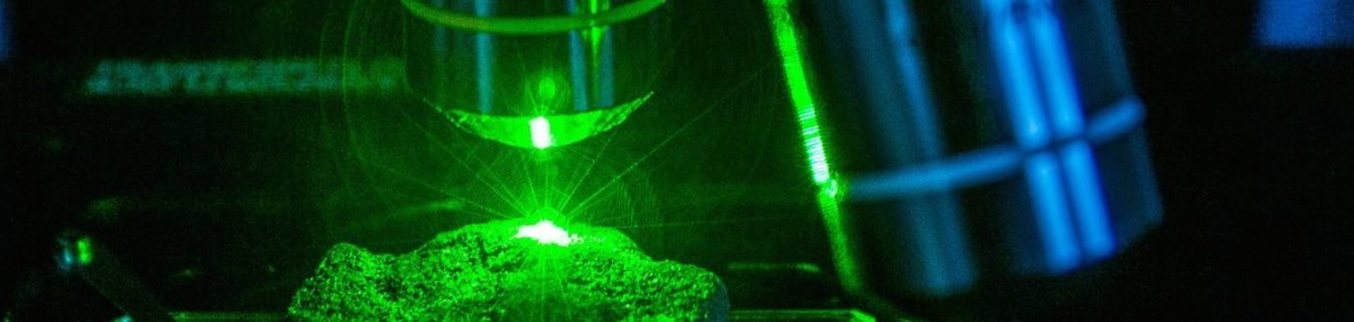
Laser Raman Microspectroscopy
Laser Raman microspectroscopy makes it possible to identify and in some cases quantitatively analyse the covalently bound chemical species in liquid inclusions.
The Dilor LabRAM instrument in Leoben works as follows: A laser beam is focused by an Olympus BX 40 microscope on the liquid inclusion of interest. The 50x and 100x magnification objectives, combined with a confocal optical arrangement, allow spatial resolution on the order of a cubic micrometre. This means that the laser can often be focused on individual phases in multiphase inclusions. Our LabRAM has two lasers: a 100 mW frequency-doubled Nd-YAG with 532 nm wavelength (green) and a less powerful He-Ne laser with 613 nm (red). With the high-energy green laser, the detection limits are lower, but the red laser is preferable if the samples to be analysed are fluorescent. Due to the interaction of the incident laser light with the molecular bonds in the target species, part of the incident light is scattered by the "Raman effect", emitting light with a frequency that is shifted from that of the laser and is characteristic of the vibrational mode and energy of the bond. Some of the scattered light is collected by the microscope and focused onto a diffraction grating. The grating selects the desired region of the Raman spectrum and reflects it onto a Peltier-cooled CCD matrix detector. The resulting spectrum (intensity versus Raman-shifted frequency) is displayed on a computer monitor for further processing and interpretation. Measurements can also be made at controlled temperatures between -190 and 600 degrees Celsius when using the Linkam THMSG 600 heating-cooling stage described above.
Further information:
Details of the application of this method to liquid inclusions can be found in:
Dubessy et al. (1989): Advances in C-O-H-N-S fluid geochemistry based on micro-Raman spectrometric analysis of fluid inclusions. European Journal of Mineralogy 1: 517-534.
Hydrothermal lab
For hydrothermal experiments up to conditions of approx. 800 C and 10 kbar, 10 externally heated, cold-sealing pressure vessels are available. Argon is used as the pressure medium. Liquid inclusions are synthesised in pre-crushed quartz sealed in gold capsules with liquid components of known concentrations. Liquid inclusions are produced in this laboratory to calibrate the temperatures of phase transitions observed in natural liquid inclusions and to provide calibration standards for analytical methods.
